Ever wondered how deadly snakes evolved their fangs? The answer lies in particular microscopic features of their teeth, research led by Flinders University and the South Australian Museum suggests.
“It’s always been a mystery why fangs have evolved so many times in snakes, but rarely in other reptiles. Our study answers this, showing how easy it is for normal snake teeth to turn into hypodermic needles,” says lead author Dr Alessandro Palci, from Flinders University.
Of almost 4,000 species of snakes alive today, about 600 of them are considered ‘medically significant’ to humans, meaning that if you get bitten you are very likely to require a visit to the nearest hospital for treatment.
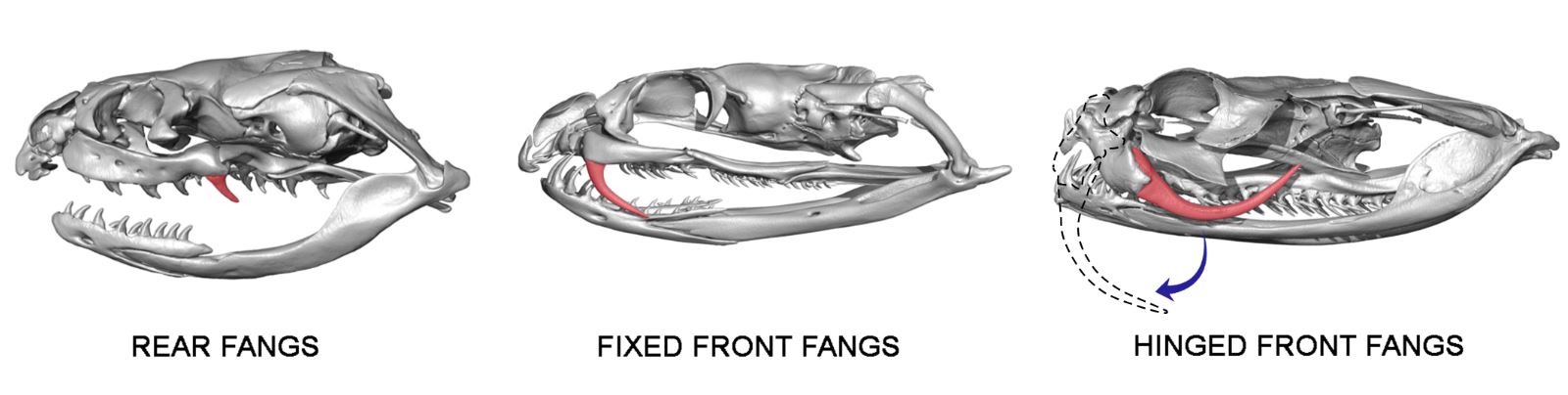
Venom fangs are modified teeth that are grooved and larger than other nearby teeth. They can be located at the back or at the front of the mouth, where they can be fixed or hinged (i.e. they can fold backwards).
Australian and overseas researchers used high-tech modelling, fossils, and hours of microscope observations to reveal that snakes possess tiny infoldings, or wrinkles, at the base of the teeth. These infoldings might help teeth attach more firmly to the jaw. In venomous snakes, one of these wrinkles becomes deeper and extends all the way to the tooth tip, thus producing a venom groove and a fang.
“Our work also highlights the opportunism and efficiency of evolution. Wrinkles which helped attach teeth to the jaw were repurposed to help inject venom,” says co-author Matthew Flinders Professor Michael Lee (Flinders University and South Australian Museum).
The paper, Plicidentine and the repeated origins of snake venom fangs (2021) by Alessandro Palci, Aaron RH LeBlanc, Olga Panagiotopoulou, Silke GC Cleuren, Hyab Mehari Abraha, Mark N Hutchinson, Alistair R Evans, Michael W Caldwell and Michael SY Lee has been published in Proceedings of the Royal Society B 288: 20211391. https://doi.org/10.1098/rspb.2021.1391
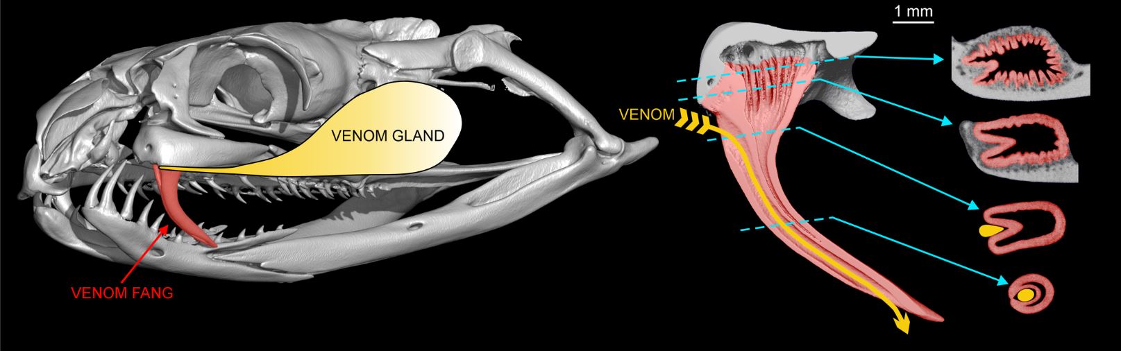
The venomous teeth of modern snakes share several common characteristics, including a venom-delivering tube or groove made up of a type of tissue called plicidentine. Plicidentine is
widespread among snakes and is key to understanding how snakes could repeatedly evolve their fangs.
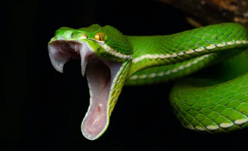
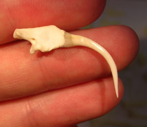
“A snake’s venom fang is an iconic example because it’s a tooth that’s been modified into a venom-injecting syringe,” said co-author Aaron LeBlanc, previously from the University of Alberta, Canada and now a Marie Curie Postdoctoral fellow at King’s College London.
“Our closer look into the development and evolution of snake teeth tells us how this might have happened through gradual, piecemeal changes,” he says.
“It reveals how mutations and subtle changes in tooth shape and size can slowly modify a tooth from a grasping and puncturing structure into a new tooth type that injects venom.”
Acknowledgements: Researchers received funding from an NSERC Postdoctoral Fellowship, NSERC Discovery Grant, Australian Research Council Discovery Program and University of Alberta grants.
Read more in The Conversation by Alessandro Palci (Research Associate in Evolutionary Biology, Flinders University), and coauthors Aaron LeBlanc (Postdoctoral Fellow in Vertebrate Palaeontology, King’s College London) and Olga Panagiotopoulou (Senior lecturer, Monash University).