Known initially as machines or contrivances, and later as manikins, phantoms, robots or dummies, ingenious models of human anatomy have been used for ‘hands-on’ medical teaching for hundreds of years.
Professor Harry Owen of the School of Medicine at Flinders University has been using high-tech mannequins and models to teach medical students the elements of anatomy and to learn a range of medical and surgical procedures for the past 20 years. His interest in the technology harnessed to teach students about the human body over the centuries has led him to write an illustrated history, Simulation in Health Care Education.
Professor Owen says there has been a strong resurgence in the past 25 years in the use of simulation in the early stages of medical training. He says modern simulators often use state-of-the-art technology that enables a range of conditions and symptoms to be simulated and provides students with realistic feedback.
“Medical schools are once again realising the benefits of allowing students to learn the basics of risky and invasive procedures without putting real patients at risk,” Professor Owen says.
“Such simulation has the added benefit of training students to treat conditions or diseases that they may never see in live patients during their training.
“However, in medical training generally, patients are still used by novices who are more likely to make mistakes than trained staff.
“Today we can identify patients who have been injured or die, and continue to be harmed, because simulation has not been adopted in healthcare as it has in fields like aviation.”
Professor Owen, a clinician, researcher and teacher in the Department of Anaesthesia and Pain Medicine, said the inventors and manufacturers of models and simulators have displayed extraordinary imagination and ingenuity in creating their imitations of human anatomy and physiology.
He says that while simple medical teaching models date back as far as Babylon, the golden age for medical modelling began in post-Renaissance Europe, driven by growing anatomical and scientific knowledge.
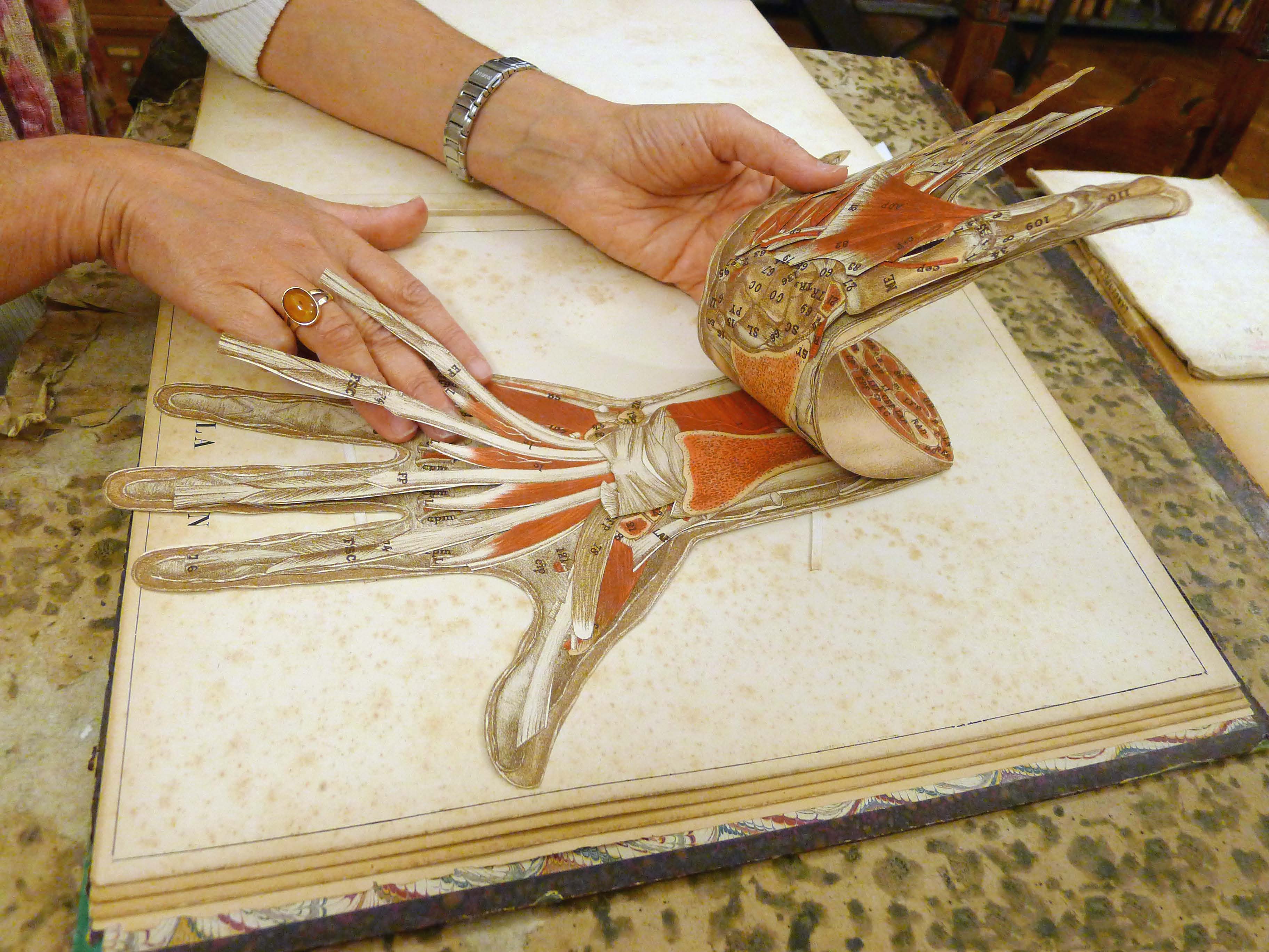
The illustrations in the book reveal the painstaking detail of models of bodies and body parts initially made from materials that included paper, papier-mâché, metal, plaster and wood. In the 17th and 18th centuries, wax became the favoured material and, apart from the lack of blood, achieved a remarkable, if sometimes gruesome, level of veracity.
Professor Owen said one wax model, a replica of a woman dissected following her death in early pregnancy, was so accurate in its modelling of the internal organs that later generations of doctors were able to identify the cardiac condition, unknown at the time of her death, that had killed her.
In the 19th century, controversy grew over whether obstetric operations should be practised using cadavers or artificial simulators. Professor Owen said that at a time before the value of handwashing was realised, a tragic wave of infant mortality ensued when medical students assisted in deliveries after using actual corpses of women and children for instruction.
The widespread use of simulators in medical education declined in the 20th century, a slump that Professor Owen attributes to the advent of large university teaching hospitals, which presented medical students with a pool of live subjects, often poor patients who had little say in their participation.
Now, he says, the dummies are back, and better than ever.
Simulation in Health Care Education: An Extensive History is published by Springer.
RELATED STORY: Flinders has introduced a network of digitally linked high-tech medical devices to revolutionise medical training in regional and rural areas. Four Anatomage tables will allow staff and students in Darwin, Mount Gambier and Renmark to link to expertise in Adelaide in real time, with all participants seeing the identical simulated anatomical procedure. Go here to read more.