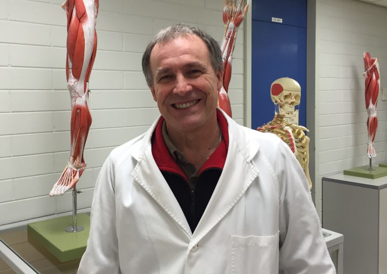
 Thousands of Flinders students owe much of their knowledge of the human body to the School of Medicine’s Anatomy Museum and to its custodian Greg Souter, who retires this week after 35 years.
Mr Souter came to Flinders as a technician in November 1981 to assist the Museum’s inaugural manager, Carlos Kordjian, and took over the Senior Technical Officer role in 2001.
The Museum, located in the Flinders Medical Centre, exists in tandem with the University’s dissection facility.
“Museum” usually suggests a static display, but the hundreds of models and specimens that make up the Flinders collection are used in different configurations for a range of practicals in anatomy.
“It doesn’t just sit here: much of what you see gets transported into our practical teaching area for particular classes,” Mr Souter says.
While the chief users are medical students, Mr Souter says five other main courses – optometry, speech pathology, paramedics, body systems (a range of medical science groups) and physiotherapy and occupational therapy – make use of the unique resource.
The main part of the museum contains a range of exhibits to familiarise students with the basics of normal human anatomy, while a smaller, separate collection exhibits a range of pathologies and abnormalities.
Mr Souter and his assistant are also in charge of storing human cadavers that have been donated to Adelaide’s medical schools, and assisting in dissection practicals with students.
The cadavers, from the joint donation program based at the University of Adelaide, come to Flinders after embalming and are stored in the cold rooms for progressive dissection over two to four years.
The medical course at Flinders includes five weeks of dissection in first year and 12 weeks in second year.
“This University is one of the few that has a full dissection course for medical students,” Mr Souter says.
“There’s a lot of good technology – we have the digital Anatomage Table here now – but it supplements the bodies, it doesn’t replace them.”
Other developing technology, in the form of digital screens that allow computer and camera images to be broadcast and made available on line, has helped in keeping up with ever-growing numbers of students, Mr Souter says. Video conferencing also allows teaching of students in remote locations, notably the Darwin-based Northern Territory Medical Program.
Mr Souter says while models present a single stylised version of musculature, nerves and blood vessels, dissection practicals, along with access to the photographs and specimens from bodies that the Museum provides, enable students to see the range of variations between individuals.
Part of Mr Souter’s highly specialised skills includes taking sections from bodies, which are then painstakingly labelled and then “potted” – preserved in formaldehyde solution or liquid paraffin in Perspex containers as sources of reference.
Familiarity with “slices” of the body is particularly important to trainee doctors who will use imaging technology, Mr Souter says.
“Being able to see what something looks like in section, particularly through the head, neck or chest, makes it much easier to interpret your scans,” he says.
Mr Souter said the Museum is by no means all his own work – in addition to the work of his predecessor, he says the departments of Medical Illustration and Media and Biomedical Engineering at FMC have made major contributions to the displays.
Mr Souter says his legacy already has a life of its own.
“I’ve got to the stage where I and members of my family are being treated by doctors who are graduates of the medical course here – I like to think that they know their anatomy.”